facebook-img

‘ANOREXIC’ DOG HAD ACTUALLY SWALLOWED SIX-INCH STICK

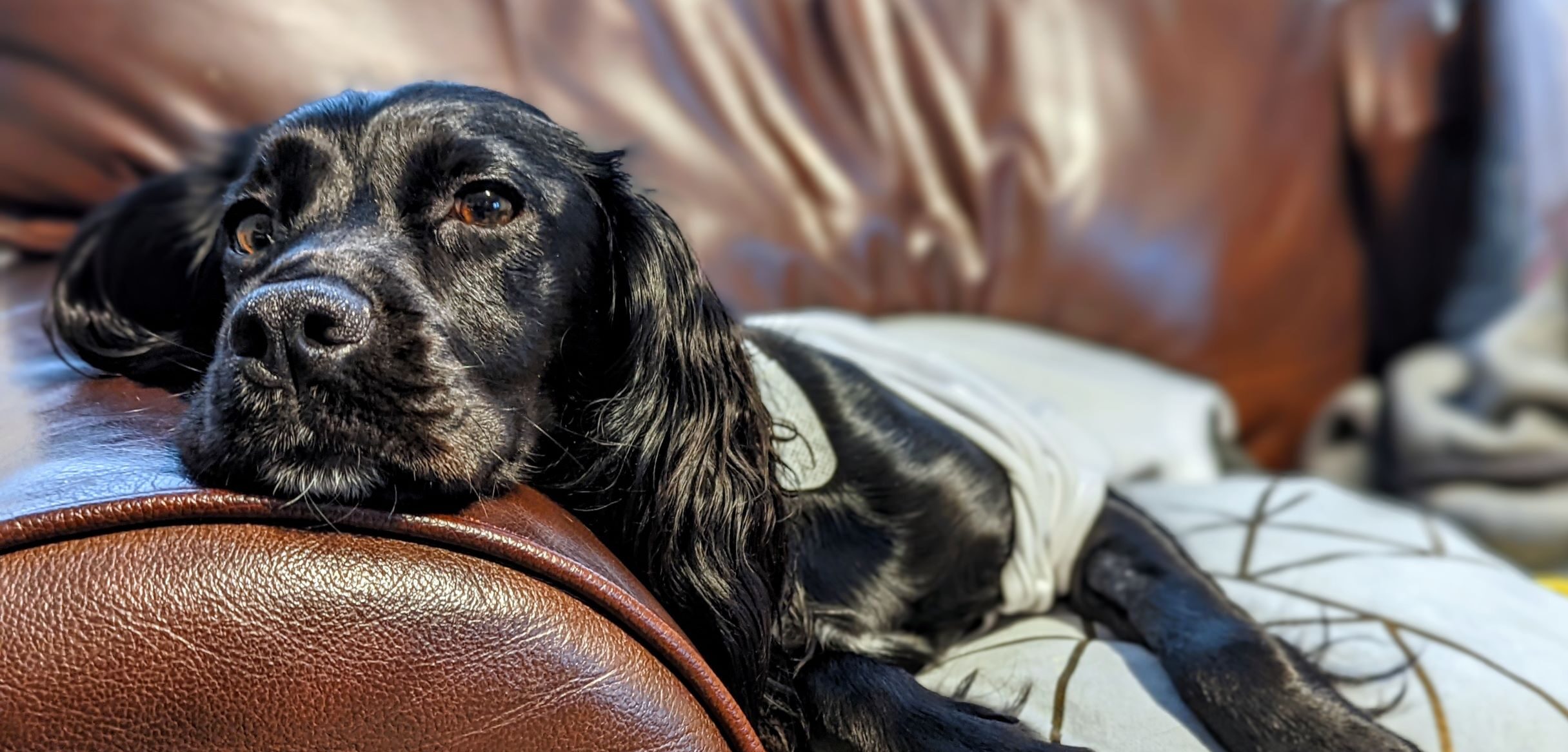
An ‘anorexic’ dog’s refusal to eat was soon explained when the one-year-old Cocker Spaniel was examined by our industry-leading multidisciplinary team. 

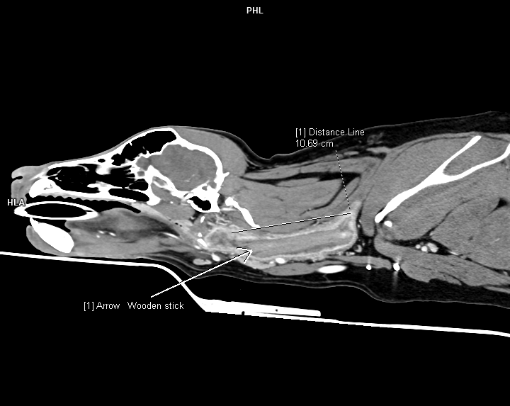
A CT scan carried out here at Willows revealed a six-inch stick had speared through the young dog’s stomach, abdominal cavity and into her pelvic canal.  

The dog, called Susan, underwent emergency surgery with our team to remove the stick and part of her stomach, and has since made a full recovery, much to the relief of worried owner Will Pool, from Wellington, Shropshire.  

Will said: “A month before her operation, Susan had almost completely stopped eating, didn’t want to play, and she struggled going downstairs and jumping up.

“We took her to our local Vets who prescribed a course of antibiotics, and she was fine again for a while.

“Then when the problems returned for a second time the Vet thought it could be steroid responsive meningitis and referred us to Willows for a Specialist diagnosis.

“We were mortified when we were given the diagnosis. We weren’t expecting her to need an operation and we certainly weren’t expecting it to be caused by having a stick inside of her.

“The Vet said the stick had been inside her for at least a few weeks, however we wonder if it might have been months because she’d been so reluctant to eat and really lethargic for so long.

“Naturally we were incredibly worried while she was having her operation especially as she was so frail from not eating so well.

“However, we were kept up to date on her progress throughout her stay and the vet always let us know before making any decisions, so we felt very well informed throughout.

“The Vets did a great job of looking after Susan and we can’t thank them enough and we would absolutely recommend Willows to anyone.

“Susan eats properly now, which she’d never really done before, and is far more energetic. We know that, without the Specialist scans and subsequent surgery, things could have ended very differently.”

Our unique multidisciplinary approach to care meant several specialists across a range of disciplines were able to work together to effectively diagnose and treat Susan’s complex condition, something we pride ourselves on greatly.

Laura Bree, an Internal Medicine Specialist here at Willows, carried out the initial investigations of Susan’s elevated temperature, lethargy and anorexia, and said: “We carried out blood and urine tests along with CT imaging of the chest and abdomen.

“It was after the CT scan of her abdomen that we received a surprise – a foreign body, consistent with a stick, was found and Susan was transferred to my colleague Will Robinson for surgery to remove it.”

Soft tissue specialist Will added: “During surgery, a long piece of stick was removed along with a small section of her stomach.

“The suspicion is that at some point Susan ate the stick which then perforated her stomach wall and travelled to the pelvic canal.

“There was also evidence of an infection in the abdomen, so she was started on a course of antibiotics after appropriate samples were taken.

“Susan was then sent for recovery in our intensive care unit under the supervision of our specialist-led emergency and critical care team.

“She recovered extremely well, and we are very pleased to report that she is now back to her old self.

“It is thanks to our multidisciplinary approach that Susan’s care involved a number of specialists across a wide range of disciplines including diagnostic imaging, internal medicine, soft tissue surgery and critical care who all worked together to diagnosis and treat this complex condition.”

PIG’S CORNEA USED IN EXPERT SURGERY TO STOP BELOVED BULLDOG GOING BLIND

A pet owner has spoken of their delight after one of our brilliant Ophthalmology team used a pig’s cornea to save a young bulldog from becoming blind.

Three-year-old Marley had already lost his left eye after suffering a deep corneal ulcer which had perforated the eyeball.

So, when he was diagnosed with an identical problem in the right eye, his owners Michael and Carol Bradley, from Walsall, feared he would completely lose his sight.

However, Alberto Palella Gomez, a Clinician in Veterinary Ophthalmology here at Willows had a plan to ensure Marley would keep his vision.

CORNEAL ULCER

Alberto explained: “Marley had a right-sided deep corneal ulcer which, in the absence of other ocular abnormalities, probably started as superficial defect and progressed to a deep corneal ulcer due to bacterial contamination.

“Marley had previously undergone right-sided corneal surgery at his referring veterinary practice where they had performed a conjunctival pedicle graft but unfortunately that had failed. They had also removed his left eye, so Marley came to us at serious risk of another perforation, and was in danger of losing his remaining eye.”

Fortunately for Marley, we are an internationally-renowned centre of excellence for Ophthalmology cases and our expert team and state-of-the-art equipment enables us to regularly treat the most complicated corneal cases, which include corneal grafting from donated tissue.

This tissue actually comes from deceased pigs’ corneas. These corneas are bio-engineered for use in repairing corneal ulcers such as Marley’s and have been used extensively in veterinary and human medicine.

EXCELLENT VISION

Alberto said: “We perform corneal surgery here at Willows on a daily basis and corneal grafting is one of these such procedures.

“We successfully performed a right-sided corneal transplant to repair the defect. Marley recovered really well from surgery and not only was he able to keep his remaining eye, he has also regained excellent vision in that eye as the cornea became almost completely transparent.”

Delighted owner Michael said: “We can’t praise Alberto and Willows enough. We’d absolutely recommend them to other pet owners. Marley’s right eye is absolutely fine again now. He’s not on any medication at all and he’s doing really well. We couldn’t be more grateful.”

To find out more about Willows’ Specialist Ophthalmology service click here.

WILLOWS PRAISED FOR LIFESAVING ‘MIRACLE’ TREATMENT

A grateful dog owner has paid a glowing tribute to the team here at Willows who saved her young Labrador’s life.

Pet owner Amanda Coleman, from Leicestershire, feared the worst throughout a dramatic four-month fight for life for three-year-old Poppy, which began when the young lab was speared in the neck by a stick.

Incredibly, four inches of the stick remained lodged in her swollen neck for more than six weeks and went unnoticed, despite several visits to local vets.

Amanda said it was only after Poppy was referred to our small animal specialist Erika Villedieu here at Willows, that the problem was finally identified.

Amanda recalled the traumatic experience, saying: “We first realised something was wrong when Poppy started coughing and gagging on her night-time walk.

“She was bleeding from the back of her mouth and gagging as if she had something stuck in her throat.

“We went to our local emergency vets who stabilised Poppy and treated the bleeding, however they were unable to see that a piece of the stick was embedded in Poppy’s neck with use of X-rays alone.  

“Four days later, Poppy came home from the emergency vet on a course of steroids with a nasogastric feeding tube but as the steroid dose was reduced over time, she became really poorly and our vet referred her to Willows.

“It was there that Poppy had her first CT scan and Erika found a four-inch piece of stick still embedded in her neck more than six weeks after her accident.

“At first, we were stunned that Poppy had been walking around with this stick still inside without anyone realizing. I was so relieved something had been found, though, and Poppy could have the surgery straight away.”

Erika operated immediately to remove the stick but said surgery was not the simple solution that was expected.

She explained: “The CT scan carried out by Willows world-leading Specialist Radiology team confirmed a four-inch piece of wooden stick had lodged itself in Poppy’s neck and was causing an abscess.

“The wooden stick was removed surgically and one of Poppy’s salivary glands also had to be removed as it had been damaged by the stick.

“Poppy received a long course of antibiotics after surgery however, despite her neck swelling having resolved, Poppy continued to experience heavy drooling, attempts to swallow and discomfort.”

Amanda added: “Poppy was withdrawn, lethargic, drooling continually, not interested in her food or anything else, and wouldn’t walk anywhere.

“A month later, Poppy was still not getting any better so we had a difficult conversation with Erika as we felt Poppy’s quality of life was being so badly affected and she was slowly starving herself.”

Erika said: “Poppy’s condition was unusual given that the wooden stick had been removed and the swelling had resolved, and so we carried out a second CT scan to further understand Poppy’s condition.

“This confirmed the abscess was resolved and that the wooden stick had been fully removed, however all of Poppy’s salivary glands appeared slightly bigger than normal.

“A presumed diagnosis of sialadenosis (swelling of the salivary glands) was made and Poppy was started on phenobarbitone, an anti-epileptic medication.

“Happily, Poppy’s signs completely resolved within two days of starting the medication and she is now back to her very lively self, enjoying her walks.”

Owner Amanda said the relief was overwhelming and praised Erika and the team here at Willows for performing “a miracle” on the family’s beloved pet.

Amanda said: “Throughout the entire four months we worried constantly. Poppy was never left alone – she even came to work with me – and we had numerous visits to our local vets in between the ones to Willows.

“We honestly thought we would lose Poppy after all she had been through.

“It was a long road to recovery for Poppy and it would never have been possible if it was not for Erika, and the team at Willows. We can’t thank them all enough for what they achieved.

“Poppy is back to her normal boisterous, silly, bouncy self. Before the accident, we always used to say that Poppy was hyperactive and when I picked her up from Willows after the second CT scan and saw her bouncing and dragging Erika across the waiting room to get to me, I knew we had our crazy girl back!

“I couldn’t stop smiling, and couldn’t thank Erika enough. She was amazing and performed nothing short of a miracle to save Poppy’s life.”

WILLOWS AT FOREFRONT OF TOTAL KNEE REPLACEMENT CARE IN UK AND EUROPE

Our top team of highly-experienced Orthopaedic Specialists are proudly leading the way across the UK and Europe for Total Knee Replacement (TKR) surgery in dogs.

We now boast an unrivalled service for Total Knee Replacement surgery, with Specialists Alexis Bilmont, Stephen Clarke and Toby Gemmill all recognised as certified TKR surgeons.

Toby, who is managing director here at Willows, said exam successes of Alexis and Stephen meant our hospital is now operating at the pinnacle of TKR care – both at home and abroad.

Toby said: “We are delighted Stephen and Alexis have now completed their training to take the number of TKR-certified surgeons at Willows up to three. This number truly puts us at the forefront of this field across the UK and Europe.

“TKR is a procedure in which a painful or poorly-functioning stifle joint is replaced with a metal and plastic prosthesis.

“The procedures can be very successful in reducing discomfort and improving limb function.

“Surgery is primarily performed in medium and large dogs with persistently painful knees, which do not respond well to medical management – most commonly in cases of severe knee osteoarthritis.”

Willows has been a long-standing pioneer of TKR surgery and hip replacement surgery, with Toby excited to now see that service now ascend to an even higher level.

He added: “We have been performing total knee replacements and hip replacements in dogs at Willows for more than 10 years, and have seen many dogs go from being chronically painful to regaining excellent function and returning to full activity.

“It has been incredibly satisfying to be able to help these dogs back to enjoying such a high quality of life.

“We’re now looking forward to our expanded service ensuring that we can help even more dogs with sore knees to become pain free and remain active.”

BIG THUMBS-UP FOR SIGN LANGUAGE VET NURSE EMMA

A Student Vet Nurse (SVN) here at Willows has recently played a vital part in a major operation to cure a dog’s lameness.

While SVN Emma Paul was not directly involved in the surgery to repair a damaged cranial cruciate ligament, her role was still essential to proceedings, as she used her sign language skills to communicate throughout with the 10-year-old cocker spaniel’s owner, who is deaf.

Emma explained: “Communication was initially difficult between our client and clinicians, so I was asked if I could help with explaining the planned treatment and operation.

“I have been signing for as long as I can remember as my older brother is deaf and I grew up going to the BID Deaf Cultural Centre in Birmingham, so was delighted to help.

“The owner was really happy when I went out and introduced myself. He was so relieved that there was someone who could understand him and explain all the important information to him.

“It was also extremely rewarding for me to be able to use my skills to help, as I know my brother struggled with going to places and not having someone there who could communicate. It was very frustrating for him. 

“The owner has requested I interpret for him when he and his pet come in again for future consultations and I have since used my sign language to help one of his friends who is also deaf.

“I’m so glad my sign language skills were able to help with this case and I will hopefully be able to assist with clients who are members of the deaf community in the future.” 

The further good news is that the cocker spaniel’s operation went well, as Kinley Smith, our RCVS and European specialist in small animal surgery, successfully repaired the CCL damage.

Dr Smith said: “Emma’s sign language skills were a huge help in this case and ensured the owner was kept fully informed throughout.

“We’re very conscious that having a sick or injured pet can be a stressful and anxious time for owners, so we always strive to communicate clearly and openly with them about the way forward.

“In this case, it was proving problematic but Emma really made the difference, not just for the owner, for all of us.”

ACE TREATMENT AS BOXER DOG LEFT ON THE ROPES AFTER SWALLOWING TENNIS BALL

A romp in the park turned into a race to the vets after a young boxer dog swallowed a tennis ball which lodged dangerously in her stomach.

Two-year-old Bonnie was rushed to us for emergency specialist treatment after swallowing a miniature tennis ball when playing with it on one of her daily walks.

However, after being left on the ropes, the boxer has now bounced back to her best following our multidisciplinary approach, which ultimately involved passing a camera through the mouth, down the oesophagus and into the stomach to retrieve the small ball.

Prior to the successful outcome, owner Dr Laura May, a consultant anaesthetist at University Hospital Coventry and Warwickshire, said her medical knowledge meant she knew how much danger the family’s beloved Bonnie had been in.

Dr May said: “I was very concerned about the potential consequences when a chest x-ray revealed the ball was in the stomach.

“Due to its size, I assumed the worst and thought it would need to be surgically removed, so I was extremely worried about the risks both intra and postoperatively.”

However, detailed discussions with our specialist-trained emergency and critical care clinician Fernanda Camacho eased her fears and a treatment plan was put in place.

Dr May added: “I was fully informed and involved in the decision-making as to how to proceed. We talked through options, from induced vomiting, watch and wait, to surgery, and decided on a step-by-step plan from least invasive with surgery a last resort.”

Fernanda said her first move was to try to induce vomiting, explaining: “I gave Bonnie an injection of apomorphine and she brought up her breakfast but, unfortunately, no ball.

“An X-ray confirmed the location of the ball was still present within Bonnie’s stomach. I was concerned the ball may cause inflammation of the stomach lining (gastritis) or obstruction in her intestine. So, in order to avoid an operation to remove the ball, Bonnie underwent endoscopy under an anaesthetic.”

Our European and RCVS internal medicine specialist Isuru Gajanayake then carried out the endoscopy, saying: “An endoscopy involves passing a flexible camera through the mouth, down the oesophagus and into the stomach.

“Using the camera, I was able to locate the ball and then using special forceps, which are guided through a channel inside the endoscope, I was able to secure the ball.

“Bonnie made a very uneventful recovery and was allowed home and is now doing very well.”

Dr May was full of praise for the Willows team, saying: “Bonnie bounced back very quickly and was back to normal the following day.

“I would most definitely recommend Willows to other pet owners. They were really professional but friendly, and they always took the time to listen and fully explain what was required.

“I felt she was in the best place and was so relieved they were able to remove the ball endoscopically. They looked after Bonnie so well that she still enjoys going there!”

CAT’S HAIR-RAISING MYSTERY SOLVED BY WILLOWS

Our expert team here at Willows ensured a family cat kept all of its nine lives by carrying out emergency surgery after we discovered the pet had ingested almost 50 hairbands.

Berry, a two-year-old domestic shorthair, was referred to us when her owner reported the cat had been vomiting and retching.

Our team took graphic x-rays which highlighted the problem – a tangled mass of elasticated hairbands lodged between Berry’s oesophagus and stomach.

Our rotating intern Jo McKendry explained: “Berry arrived having suffered acute vomiting the night before and she now wasn’t eating or drinking. 

“She behaved like she was trying to vomit up a fur ball but was struggling to bring it up.

“She was clearly dehydrated and when I examined her, I felt a large sausage-style structure in her cranial abdomen which was confirmed by x-rays. 

“The images showed a foreign body extending from Berry’s stomach into her distal oesophagus – and it appeared to be a mass of hairbands.  

“I performed a gastrotomy to access and remove the bands, and Berry was allowed home the following day. 

“She was quickly back to normal, showed a massive increase in appetite and was soon putting on weight.” 

Owner Paul Spraggett, from Stratford, Warwickshire, was left stunned when our team told him the cause of Berry’s condition was down to her habit of snaffling the elasticated accessories.

After leaving the beloved family pet to undergo emergency surgery, Paul said he immediately went home to warn his wife and two daughters about the dangers of leaving hairbands lying around.  

He said: “None of us had ever thought about the risks that hairbands and similar everyday objects could pose for our four cats. 

“We were really shocked to find out that was why Berry had been so sick and that’s why hairbands are now banned from anywhere the cats might go in the house. 

“Willows told us they’d had other similar cases in the past, although not involving anything like the amount that Berry had swallowed.

“Jo and the team at Willows were absolutely fantastic from start to finish. They explained everything to us very clearly and thoroughly and kept us in touch every step of the way.

“We’re so grateful to have our lovely cat back and she’s certainly loving being back, too. She’s racing around with her brother and has really come on and grown since the operation. 

“Now she has much more of an appetite and that’s convinced us that she must have been snaffling these hairbands over a long period of time, maybe even 18 months. 

“We think they gradually accumulated, filling her stomach and that’s why she could only eat her food in small portions. 

“Finally, her stomach was so full it made her ill and we ended up at Willows to discover why.”    

EMERGENCY PACEMAKER CURES CONSTANTLY COLLAPSING CANINE

A constantly collapsing canine has undergone emergency life-saving surgery here at Willows to cure his dangerously low heartbeat.

Seven-year-old German Shepherd, Dexter, was suffering from a condition called Persistent Atrial Standstill (PAS), which was causing him to crash to the floor every few minutes, leaving his devoted owners fearing they might lose their beloved pet.

However, Dexter was swiftly referred to us as an emergency out-of-hours case for expert care.

RCVS and European specialist in small animal cardiology Fabio Sarcinella decided to operate on the same night Dexter was admitted because the dog’s condition was so serious.

Fabio said: “Dexter was collapsing every few minutes, even with minimal exercise, as his heart rate continued to slow down alarmingly.

“We carried out an electrocardiogram (ECG) which confirmed the diagnosis of Persistent Atrial Standstill (PAS), which occurs when the top chamber of the heart stops working and results in a very low heart rate.

“In Dexter’s case, his heart rate could go as low as 36 beats per minute, causing his collapsing episodes. The normal heart rate is between 80 and 120 beats per minute.

“In the circumstances, the only available treatment for Dexter was the fitting of an emergency pacemaker, which is a small device that sends electrical impulses to the heart to keep it beating regularly and not too slowly.

“Those electrical impulses reach the heart through a wire (pacemaker lead) that is inserted into the heart via one of the body vessels.”

Dexter recovered well from surgery and was allowed home just two days later, much to the delight and relief of his concerned owner Daniel Pickup, from Boston in Lincolnshire.

He said: “Willows gave us hope in a really difficult time – just what any pet owner would want – and they have quite literally given Dexter his life back. 

“Although the diagnosis was a shock, the opportunity to give him a good life again with a pacemaker was not a hard decision to make.

“The prognosis without treatment was not positive at all and we weren’t prepared to lose him at such a young age.

“We knew the operation came with risks and that there could be complications but we felt as if we were in safe hands. Fabio was extremely knowledgeable and reassuring throughout the whole process.

“We have since returned to see Fabio for Dexter’s first post-operative check and the results show what a success it has been.”

MULTIDISCIPLINARY APPROACH HELPS TO SOLVE SHUNT CASE

A border terrier has undergone lifesaving surgery thanks to a multidisciplinary approach by the specialist-led team here at Willows.

Four-year-old Henry was referred to us after his concerned owners reported his lack of energy and general lethargy, including not even being able to go out for his usual daily walks.

Mark Dunning, head of internal medicine and an RCVS and European Specialist in small animal internal medicine, first saw Henry and found a shunting vessel on an abdominal ultrasound scan which had been performed by our specialist-led imaging team.

Following the discovery, Henry was then seen by Will Robinson, RCVS and European Specialist in small animal surgery, for management of the portosystemic shunt, a condition in which an abnormal blood vessel allows blood to bypass, or shunt, around the liver.

Will said: “A liver shunt occurs when an abnormal connection exists between the portal system, which drains the intestines and some other abdominal organs, and the vena cava or another major vein.

“These conditions can be life-limiting for those dogs affected and, as seen with Henry, can have a severe effect on their day-to-day capabilities.

“Evidence supports that dogs have more of a normal life expectancy with surgical management, compared with medical management, so his owners elected to proceed with surgery.”

Henry was anaesthetised for his surgery by our specialist-led anaesthesia team and taken to theatre, where Will carried out surgery to locate the abnormal vessel which was then closed with a permanent suture.

Will said: “The surgery was successful and Henry recovered well from his procedure. We are pleased to report that he is doing really well at home, is off all medications and his owner’s report he is almost like a puppy again!”

WILLOWS TEAM SAVES LIFE OF PARALYSED PUPPY MYLO

A paralysed puppy that was suffering from a rare life-threatening condition has made a spectacular recovery after 11 days in intensive care and dedicated physiotherapy as part of a truly multidisciplinary approach here at Willows.

Mylo, a two-month-old Sproodle, was admitted to us as an emergency case after becoming paralysed, which included losing the ability to bark, yelp and whimper.

Our Neurologist Resident Rob Clark carried out an examination on Mylo which pointed towards a condition called ‘immune-mediated polyradiculoneuritis’, which was confirmed by a series of complex neurological diagnostic tests.  

Rob explained: “Mylo’s own immune system was attacking his nervous system, leaving him unable to move any of his legs or even lift his head.

“I had to prepare Mylo’s owners for the worst, as polyradiculoneuritis can be life-threatening if it progresses to paralysing the nerves which control breathing.

“The other major problem was that there is no specific medical or surgical treatment for polyradiculoneuritis, so Mylo’s recovery now depended on round-the-clock intensive nursing care and physiotherapy.”

INTENSIVE CARE UNIT

Mylo was immediately admitted to our Intensive Care Unit (ICU), where he was closely monitored day and night by our dedicated team.

Rob added: “For patients such as Mylo, we have to watch and wait to see if the nerves start to heal.

Some patients deteriorate and others slowly seem to stabilise and then show signs of improvement.

“The biggest danger was that he may develop secondary complications, such as struggling to breathe or a chest infection, so the ICU team were monitoring Mylo’s vital signs 24/7 and were turning or repositioning him every four hours and propping him upright for any feeding and bathing, as well as playing and interacting with him.

“Another potential complication was that Mylo could suffer long-term mobility issues when he recovered. He was paralysed from the neck down, so he was not moving his legs at all and, as a puppy, he was still in his growing phase.

“Patients who spend so much time without moving can develop a contracture, where the muscles start to contract and pull the legs into a curled-up position, which is irreversible.

“If Mylo did develop a contracture it would mean he wouldn’t be able to walk again and puppies are especially prone to this complication as they are still growing.”

VETERINARY PHYSIOTHERAPY

This is where our expert Veterinary Physiotherapist Emma Box became key to ensuring Mylo avoided a contracture and went on to make a complete recovery.

Emma explained: “An exercise programme is essential to prevent a contracture, so I spent a long time massaging and stretching Mylo’s legs and moving his joints for him.

“Then, as Mylo started to recover, we began a course of rehabilitation. Firstly, we started the process of teaching Mylo how to hold normal positions such as sitting and lying.

“We introduced exercises which involved manually positioning Mylo into a lying or sitting position, and slowly taking support away over time.

“Initially, he didn’t have the strength to hold himself upright, but gradually you would take your hands away and he would hold himself up.

“Once he could hold these positions independently, you use further exercises to challenge his balance a little more – such as moving a treat from side to side and seeing if he can hold the sit or lie without collapsing to the side.

MULTIDISCIPLINARY

“These exercises don’t sound that strenuous but for such a tiny puppy recovering from polyradiculoneuritis they are incredibly challenging.

“Gradually were introduced to encourage him to push himself up into a standing position and try to take steps.

“When Mylo finally went home his owners also had a prescribed exercise plan to perform every four hours. They showed incredible tenacity and resilience with his aftercare and, without their efforts, I don’t think Mylo would have made the recovery he did.

“There were a few setbacks initially as we feared that Mylo had developed a contracture, but eventually, after three long months of gruelling rehabilitation at home and countless trips back to Willows, Mylo is now able to explore the exciting world like a puppy once again!”

Another successful outcome for our multidisciplinary team!

We offer specialist services in anaesthesia and analgesia, cardiology, clinical nutrition, dermatology, diagnostic imaging, emergency and critical care, internal medicine, neurology, oncology, ophthalmology, orthopaedics, soft tissue surgery and spinal surgery.